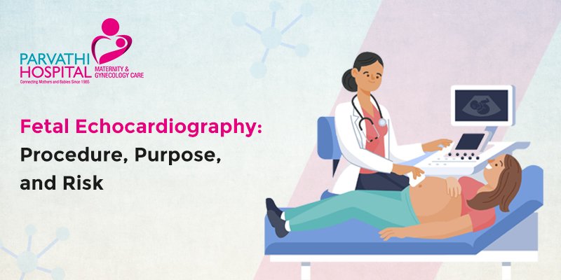
Fetal Echocardiography- Procedure, Purpose and Risk

Some expectant mothers are more likely to deliver a child with congenital heart disease or CHD. One per cent of infants are born with mild to severe heart conditions. When the CHD is discovered while the child is still in the womb, it is advantageous. Prenatal CHD diagnosis has been found to improve the pregnancy’s outcome. This is because prenatal diagnosis enables the baby to get medical and surgical intervention (if necessary) more quickly after birth and assists the expecting mother in managing the pregnancy and delivery in the event of any abnormalities. One such procedure for diagnosing fetal CHD before delivery is fetal echocardiography. For a detailed checkup, you can visit Parvati hospital. They are also known famously for having the best infertility doctor in Hyderabad.
A test called fetal echocardiography allows your doctor to view the heart of your developing kid. By employing sound waves to examine the structure of the heart, this ultrasound scan can detect a heart abnormality before your baby is born. Compared to a typical prenatal ultrasound, fetal echocardiography provides a more detailed picture of the heart of your unborn child. Guidelines for fetal echocardiography will be discussed in this article, along with preparation tips.
What is Fetal echocardiography?
A specific type of ultrasound examination called fetal echocardiography is used to examine a fetus’s heart for problems thoroughly, and abnormalities include improperly functioning heart valves, holes in the heart, narrowing arteries, irregular heartbeats, etc. Fetal echocardiography assesses the shape, structure, and functionality of the heart.
When to undergo fetal echocardiography?
Not every pregnancy involves fetal echocardiography as a standard technique. However, a simple ultrasound, a component of prenatal care, may determine if the baby’s heart has formed into all four chambers in most pregnancies. The following conditions will be recommended for doing this surgery by the obstetrician:
If the mother has any conditions that put the pregnancy in the high-risk group, such as type 1 diabetes (insulin dependent), phenylketonuria, which is an inability to break down the amino acid phenylalanine, lupus, connective tissue disease, etc.
- If a standard prenatal examination identifies any irregularities with the heart’s structure or function
- If a fetal heartbeat is found to be abnormal
- If an amniocentesis reveals a genetic or chromosomal abnormality
- If the mother drank alcohol or took drugs while pregnant
- If a heart related diseases in inherited by the family
- If the pregnant mother has a congenital cardiac condition
- If the expectant mother has already had a child with congenital heart disease
- If the expectant mother was taking any drugs that would interfere with the fetus’s normal heart development and raise the likelihood that the child would be born with congenital heart disease. Examples include medications for severe acne (like Accutane) and epilepsy.
Importance of fetal echocardiography:
Fetal echos look for congenital heart conditions that develop in the womb and may impact your baby’s heart’s structure and function. There are many pregnancy scans for expecting mothers to determine their baby’s health. For example, a more severe instance of CHD can involve missing pieces, whereas a minor case might be a hole in your baby’s heart. Examples of CHD include:
- Septal defect of the atria
- Defect in the atrioventricular septum
- An aortic coarctation
- D-transposition of the major arteries with two outlets in the right ventricle
- Anomaly of Ebstein
- Syndrome of a hypoplastic left heart
- Damaged aortic arch
- Respiratory atresia
- Only one ventricle
- Fallot tetralogy
- Abnormal pulmonary venous return in its entirety
- Atresia tricuspid
- Arteriosus Truncus
- Defect in the ventricle septum
Risks associated with echocardiography:
The potential for familial and parental anxiety, false-positive diagnoses, additional unnecessary testing and interventions, the undetermined cost-effectiveness and a priori determined risk level, as well as the potential for false-positive results, are every risk coupled with performing the procedure of fetal echocardiography, particularly in the absence of cardiac related issue finding on routine screening. Furthermore, in a thorough anatomical evaluation, most significant heart abnormalities are not overlooked.
The procedure of fetal echocardiography:
Fetal echocardiography has risks, particularly when there are no cardiac findings on routine screening. These risks include the potential for family and parental anxiety, false positives diagnoses, extra unneeded tests and actions, the unresolved cost-effectiveness and a priori identified risk level, as well as the an opportunity for false-positive results. In a complete anatomical evaluation, the most severe heart anomalies are also taken into account.
Fetal echocardiography can also be done with an endovaginal ultrasound. A tiny ultrasound transmitter is put into the vagina during this type of examination to capture photos of the heart of your unborn child. The transvaginal ultrasound is frequently performed early on and provides a clearer image.
A fetal echocardiography is normally performed by a registered cardiac sonographer (RCS), a medical expert with specialized training in operating ultrasound equipment and evaluating a baby’s heart. After the operation, a pediatric cardiologist reviews the test results. The identification and treatment of cardiac issues in newborns and young children have been the focus of this doctor’s specialized training.
How to prepare for fetal echo?
Some ultrasonography procedures benefit from a full bladder. Fetal echocardiography doesn’t require that. Since the examination could last up to two hours, you may empty your bladder beforehand. Because your doctor refers you to a specialist, providing the technician with as much information as possible is crucial. This could include information like:
- You have a cardiac defect.
- Heart abnormalities run in families.
- Clinical data Information about your issues.
Result of fetal echocardiogram:
To check for heart problems in your unborn kid, a fetal echocardiogram is performed. The development or operation of the heart may be the root of several problems. It’s also possible to see arrhythmias, or disturbances in the infant’s cardiac rhythm. When fetal echocardiography reveals that your baby’s heart has a defect, it is typically repeated to confirm the results. The pediatric cardiologist will then routinely schedule appointments with you to talk about cardiac issues and how they may affect your baby’s overall health.
Although they can be performed vaginally rather than abdominally as early as 12 weeks gestation, fetal echocardiograms are only thought to be reliable after 17 weeks. You will be informed of your baby’s best and worst options if a heart problem is found.
This aids in getting you ready for the necessary postpartum procedures. In some circumstances, newborn babies require immediate observation or surgery to stabilize them. Your doctor presents you with all of your options, educates you on the outcomes of each, and then lets you and your partner decide which course of action is best for you.
Final words:
An ultrasound test called fetal echocardiography looks at the developing baby’s heart’s structural components. This examination can find heart issues like faults or arrhythmias. Your doctor could advise getting fetal echocardiography if you have a family history of congenital heart abnormalities. It could take 45 minutes to two hours to complete the process. Usually, results are available within 24 hours.
Frequently asked questions:
A specialized ultrasound test called fetal echocardiogram is carried out during pregnancy to assess the position, size, shape, function, and rhythm of the developing baby’s heart.
When there is worry that the unborn child may have cardiac disease or is more likely to develop it, a fetal echocardiography is typically performed.
There are no hazards associated with fetal echo for either the mother or the fetus. The smallest ultrasound settings are applied.


